

ITT1588
ITT1588
ITT1588
from
$424.00
Price in reward points: 424
- Catalog: ITT1588
- Gene/Protein: EPHB6
- Product Description: Immunotag™ EphB6 Polyclonal Antibody
-
Description
-
Specifications
Antibody Specification Datasheet 
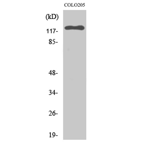
IMPORTANT NOTE This product is custom manufactured with a lead time of 3-4 weeks. Once in production, this item cannot be cancelled from an order and is not eligible for return. Target Protein EPHB6 Clonality Polyclonal Storage/Stability -20°C/1 year Application WB,IHC-p,IF,ELISA Recommended Dilution Western Blot: 1/500 - 1/2000. Immunohistochemistry: 1/100 - 1/300. Immunofluorescence: 1/200 - 1/1000. ELISA: 1/5000. Not yet tested in other applications. Concentration 1 mg/ml Reactive Species Human,Mouse,Rat Host Species Rabbit Immunogen Synthesized peptide derived from EphB6, at AA range: 830-910 Specificity EphB6 Polyclonal Antibody detects endogenous levels of EphB6 protein. Purification The antibody was affinity-purified from rabbit antiserum by affinity-chromatography using epitope-specific immunogen Form Liquid in PBS containing 50% glycerol, 0.5% BSA and 0.02% sodium azide. Gene Name EPHB6 Accession No. O15197 O08644 P0C0K7 Alternate Names EPHB6; Ephrin type-B receptor 6; HEP; Tyrosine-protein kinase-defective receptor EPH-6 Description EPH receptor B6(EPHB6) Homo sapiens This gene encodes a member of a family of transmembrane proteins that function as receptors for ephrin-B family proteins. Unlike other members of this family, the encoded protein does not contain a functional kinase domain. Activity of this protein can influence cell adhesion and migration. Expression of this gene is downregulated during tumor progression, suggesting that the protein may suppress tumor invasion and metastasis. Alternative splicing results in multiple transcript variants. [provided by RefSeq, Jul 2013], Cell Pathway/ Category Axon guidance, Protein Expression Brain, Subcellular Localization extracellular region,cytosol,plasma membrane,integral component of plasma membrane,cell surface,integral component of membrane, Protein Function domain:The protein kinase domain is predicted to be catalytically inactive. Its extracellular domain is capable of promoting cell adhesion and migration in response to low concentrations of ephrin-B2, but its cytoplasmic domain is essential for cell repulsion and inhibition of migration induced by high concentrations of ephrin-B2.,function:Kinase-defective receptor for members of the ephrin-B family. Binds to ephrin-B1 and ephrin-B2. Modulates cell adhesion and migration by exerting both positive and negative effects upon stimulation with ephrin-B2. Inhibits JNK activation, T cell receptor-induced IL-2 secretion and CD25 expression upon stimulation with ephrin-B2.,PTM:Ligand-binding increases phosphorylation on tyrosine residues. Phosphorylation on tyrosine residues is mediated by transphosphorylation by the catalytically active EPHB1 in a ligand-independent manner. Tyrosine phosphorylation of the receptor may act as a switch on the functional transition from cell adhesion/attraction to de-adhesion/repulsion.,similarity:Belongs to the protein kinase superfamily. Tyr protein kinase family. Ephrin receptor subfamily.,similarity:Contains 1 protein kinase domain.,similarity:Contains 1 SAM (sterile alpha motif) domain.,similarity:Contains 2 fibronectin type-III domains.,subunit:Interacts with CBL and EPHB1. Interacts with FYN; this interaction takes place in a ligand-independent manner.,tissue specificity:Expressed in brain. Expressed in non invasive breast carcinoma cell lines (at protein level). Strong expression in brain and pancreas, and weak expression in other tissues, such as heart, placenta, lung, liver, skeletal muscle and kidney. Expressed in breast non invasive tumors but not in metastatic lesions. Isoform 3 is expressed in cell lines of glioblastomas, anaplastic astrocytomas, gliosarcomas and astrocytomas. Isoform 3 is not detected in normal tissues., Usage For Research Use Only! Not for diagnostic or therapeutic procedures. - Reviews
| Antibody Specification | |
| Datasheet | |
| IMPORTANT NOTE | This product is custom manufactured with a lead time of 3-4 weeks. Once in production, this item cannot be cancelled from an order and is not eligible for return. |
| Target Protein | EPHB6 |
| Clonality | Polyclonal |
| Storage/Stability | -20°C/1 year |
| Application | WB,IHC-p,IF,ELISA |
| Recommended Dilution | Western Blot: 1/500 - 1/2000. Immunohistochemistry: 1/100 - 1/300. Immunofluorescence: 1/200 - 1/1000. ELISA: 1/5000. Not yet tested in other applications. |
| Concentration | 1 mg/ml |
| Reactive Species | Human,Mouse,Rat |
| Host Species | Rabbit |
| Immunogen | Synthesized peptide derived from EphB6, at AA range: 830-910 |
| Specificity | EphB6 Polyclonal Antibody detects endogenous levels of EphB6 protein. |
| Purification | The antibody was affinity-purified from rabbit antiserum by affinity-chromatography using epitope-specific immunogen |
| Form | Liquid in PBS containing 50% glycerol, 0.5% BSA and 0.02% sodium azide. |
| Gene Name | EPHB6 |
| Accession No. | O15197 O08644 P0C0K7 |
| Alternate Names | EPHB6; Ephrin type-B receptor 6; HEP; Tyrosine-protein kinase-defective receptor EPH-6 |
| Description | EPH receptor B6(EPHB6) Homo sapiens This gene encodes a member of a family of transmembrane proteins that function as receptors for ephrin-B family proteins. Unlike other members of this family, the encoded protein does not contain a functional kinase domain. Activity of this protein can influence cell adhesion and migration. Expression of this gene is downregulated during tumor progression, suggesting that the protein may suppress tumor invasion and metastasis. Alternative splicing results in multiple transcript variants. [provided by RefSeq, Jul 2013], |
| Cell Pathway/ Category | Axon guidance, |
| Protein Expression | Brain, |
| Subcellular Localization | extracellular region,cytosol,plasma membrane,integral component of plasma membrane,cell surface,integral component of membrane, |
| Protein Function | domain:The protein kinase domain is predicted to be catalytically inactive. Its extracellular domain is capable of promoting cell adhesion and migration in response to low concentrations of ephrin-B2, but its cytoplasmic domain is essential for cell repulsion and inhibition of migration induced by high concentrations of ephrin-B2.,function:Kinase-defective receptor for members of the ephrin-B family. Binds to ephrin-B1 and ephrin-B2. Modulates cell adhesion and migration by exerting both positive and negative effects upon stimulation with ephrin-B2. Inhibits JNK activation, T cell receptor-induced IL-2 secretion and CD25 expression upon stimulation with ephrin-B2.,PTM:Ligand-binding increases phosphorylation on tyrosine residues. Phosphorylation on tyrosine residues is mediated by transphosphorylation by the catalytically active EPHB1 in a ligand-independent manner. Tyrosine phosphorylation of the receptor may act as a switch on the functional transition from cell adhesion/attraction to de-adhesion/repulsion.,similarity:Belongs to the protein kinase superfamily. Tyr protein kinase family. Ephrin receptor subfamily.,similarity:Contains 1 protein kinase domain.,similarity:Contains 1 SAM (sterile alpha motif) domain.,similarity:Contains 2 fibronectin type-III domains.,subunit:Interacts with CBL and EPHB1. Interacts with FYN; this interaction takes place in a ligand-independent manner.,tissue specificity:Expressed in brain. Expressed in non invasive breast carcinoma cell lines (at protein level). Strong expression in brain and pancreas, and weak expression in other tissues, such as heart, placenta, lung, liver, skeletal muscle and kidney. Expressed in breast non invasive tumors but not in metastatic lesions. Isoform 3 is expressed in cell lines of glioblastomas, anaplastic astrocytomas, gliosarcomas and astrocytomas. Isoform 3 is not detected in normal tissues., |
| Usage | For Research Use Only! Not for diagnostic or therapeutic procedures. |