

ITA4999
ITA4999
ITA4999
from
$526.00
Price in reward points: 526
- Catalog: ITA4999
- Gene/Protein: HLA-DPA1
- Product Description: Immunotag™ HLA-DPA1 Antibody
-
Description
-
Specifications
Antibody Specification Datasheet 
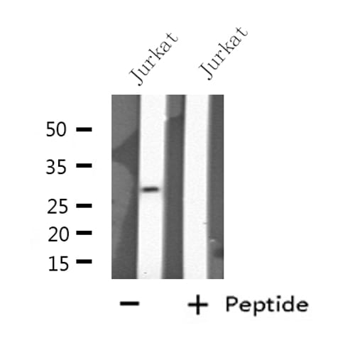
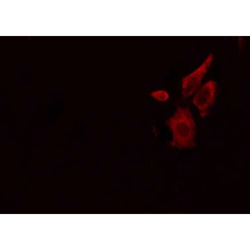
IMPORTANT NOTE This product is custom manufactured with a lead time of 3-4 weeks. Once in production, this item cannot be cancelled from an order and is not eligible for return. Target Protein HLA-DPA1 Clonality Polyclonal Storage/Stability -20°C/1 year Application WB,IF/ICC,ELISA Recommended Dilution WB 1:500~1:1000, IF/ICC 1:100-1:500 Concentration 1 mg/ml Reactive Species Human Host Species Rabbit Immunogen A synthesized peptide Specificity HLA-DPA1 Antibody detects endogenous levels of total HLA-DPA1 Purification The antiserum was purified by peptide affinity chromatography. Form Rabbit IgG in phosphate buffered saline , pH 7.4, 150mM NaCl, 0.02% sodium azide and 50% glycerol.Store at -20 °C.Stable for 12 months from date of receipt Gene Name HLA-DPA1 Accession No. P20036 Alternate Names DP(W3); DP(W4); HLA class II histocompatibility antigen DP alpha 1 chain; HLA class II histocompatibility antigen DP alpha chain; HLA DP1A; HLA DPA 1; HLA SB alpha chain; HLADP; HLADPA1; HLASB; HLASB histocompatibility type; Human MHC class II HLA SB alpha gene; Major histocompatibility complex class II DP alpha 1; MHC class II antigen; MHC class II DP3 alpha; MHC class II DPA1; MHC class II HLA DPA1 antigen; PLT1; Primed lymphocyte test 1; Description Binds peptides derived from antigens that access the endocytic route of antigen presenting cells (APC) and presents them on the cell surface for recognition by the CD4 T-cells. The peptide binding cleft accommodates peptides of 10-30 residues. The peptides presented by MHC class II molecules are generated mostly by degradation of proteins that access the endocytic route, where they are processed by lysosomal proteases and other hydrolases. Exogenous antigens that have been endocytosed by the APC are thus readily available for presentation via MHC II molecules, and for this reason this antigen presentation pathway is usually referred to as exogenous. As membrane proteins on their way to degradation in lysosomes as part of their normal turn-over are also contained in the endosomal/lysosomal compartments, exogenous antigens must compete with those derived from endogenous components. Autophagy is also a source of endogenous peptides, autophagosomes constitutively fuse with MHC class II loading compartments. In addition to APCs, other cells of the gastrointestinal tract, such as epithelial cells, express MHC class II molecules and CD74 and act as APCs, which is an unusual trait of the GI tract. To produce a MHC class II molecule that presents an antigen, three MHC class II molecules (heterodimers of an alpha and a beta chain) associate with a CD74 trimer in the ER to form a heterononamer. Soon after the entry of this complex into the endosomal/lysosomal system where antigen processing occurs, CD74 undergoes a sequential degradation by various proteases, including CTSS and CTSL, leaving a small fragment termed CLIP (class-II-associated invariant chain peptide). The removal of CLIP is facilitated by HLA-DM via direct binding to the alpha-beta-CLIP complex so that CLIP is released. HLA-DM stabilizes MHC class II molecules until primary high affinity antigenic peptides are bound. The MHC II molecule bound to a peptide is then transported to the cell membrane surface. In B-cells, the interaction between HLA-DM and MHC class II molecules is regulated by HLA-DO. Primary dendritic cells (DCs) also to express HLA-DO. Lysosomal microenvironment has been implicated in the regulation of antigen loading into MHC II molecules, increased acidification produces increased proteolysis and efficient peptide loading. Cell Pathway/ Category Primary Polyclonal Antibody Protein MW 29 KD Usage For Research Use Only! Not for diagnostic or therapeutic procedures. - Reviews
| Antibody Specification | |
| Datasheet |
|
| IMPORTANT NOTE | This product is custom manufactured with a lead time of 3-4 weeks. Once in production, this item cannot be cancelled from an order and is not eligible for return. |
| Target Protein | HLA-DPA1 |
| Clonality | Polyclonal |
| Storage/Stability | -20°C/1 year |
| Application | WB,IF/ICC,ELISA |
| Recommended Dilution | WB 1:500~1:1000, IF/ICC 1:100-1:500 |
| Concentration | 1 mg/ml |
| Reactive Species | Human |
| Host Species | Rabbit |
| Immunogen | A synthesized peptide |
| Specificity | HLA-DPA1 Antibody detects endogenous levels of total HLA-DPA1 |
| Purification | The antiserum was purified by peptide affinity chromatography. |
| Form | Rabbit IgG in phosphate buffered saline , pH 7.4, 150mM NaCl, 0.02% sodium azide and 50% glycerol.Store at -20 °C.Stable for 12 months from date of receipt |
| Gene Name | HLA-DPA1 |
| Accession No. | P20036 |
| Alternate Names | DP(W3); DP(W4); HLA class II histocompatibility antigen DP alpha 1 chain; HLA class II histocompatibility antigen DP alpha chain; HLA DP1A; HLA DPA 1; HLA SB alpha chain; HLADP; HLADPA1; HLASB; HLASB histocompatibility type; Human MHC class II HLA SB alpha gene; Major histocompatibility complex class II DP alpha 1; MHC class II antigen; MHC class II DP3 alpha; MHC class II DPA1; MHC class II HLA DPA1 antigen; PLT1; Primed lymphocyte test 1; |
| Description | Binds peptides derived from antigens that access the endocytic route of antigen presenting cells (APC) and presents them on the cell surface for recognition by the CD4 T-cells. The peptide binding cleft accommodates peptides of 10-30 residues. The peptides presented by MHC class II molecules are generated mostly by degradation of proteins that access the endocytic route, where they are processed by lysosomal proteases and other hydrolases. Exogenous antigens that have been endocytosed by the APC are thus readily available for presentation via MHC II molecules, and for this reason this antigen presentation pathway is usually referred to as exogenous. As membrane proteins on their way to degradation in lysosomes as part of their normal turn-over are also contained in the endosomal/lysosomal compartments, exogenous antigens must compete with those derived from endogenous components. Autophagy is also a source of endogenous peptides, autophagosomes constitutively fuse with MHC class II loading compartments. In addition to APCs, other cells of the gastrointestinal tract, such as epithelial cells, express MHC class II molecules and CD74 and act as APCs, which is an unusual trait of the GI tract. To produce a MHC class II molecule that presents an antigen, three MHC class II molecules (heterodimers of an alpha and a beta chain) associate with a CD74 trimer in the ER to form a heterononamer. Soon after the entry of this complex into the endosomal/lysosomal system where antigen processing occurs, CD74 undergoes a sequential degradation by various proteases, including CTSS and CTSL, leaving a small fragment termed CLIP (class-II-associated invariant chain peptide). The removal of CLIP is facilitated by HLA-DM via direct binding to the alpha-beta-CLIP complex so that CLIP is released. HLA-DM stabilizes MHC class II molecules until primary high affinity antigenic peptides are bound. The MHC II molecule bound to a peptide is then transported to the cell membrane surface. In B-cells, the interaction between HLA-DM and MHC class II molecules is regulated by HLA-DO. Primary dendritic cells (DCs) also to express HLA-DO. Lysosomal microenvironment has been implicated in the regulation of antigen loading into MHC II molecules, increased acidification produces increased proteolysis and efficient peptide loading. |
| Cell Pathway/ Category | Primary Polyclonal Antibody |
| Protein MW | 29 KD |
| Usage | For Research Use Only! Not for diagnostic or therapeutic procedures. |